Human Body Bones Diagram : Bones Of The Human Body Anatomy Physioadvisor / A human body bones diagram is usually a simplified typical pictorial illustration of an electrical circuit.
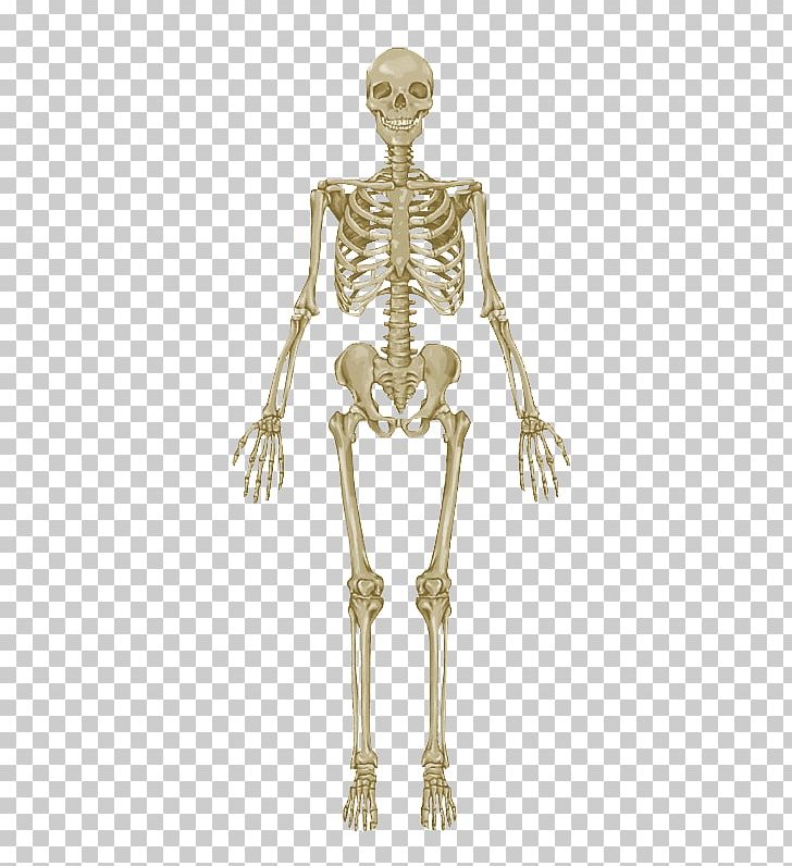
Human Body Bones Diagram : Bones Of The Human Body Anatomy Physioadvisor / A human body bones diagram is usually a simplified typical pictorial illustration of an electrical circuit.. Temporal bone occipital bone mandible humerus femur tibia calcaneus fibula ulna radius scapula clavicle scapula. Posted on may 24, 2016 by admin. Posted in bones, diagrams | tagged body skeleton, human skeletal anatomy, human skeleton, human skeleton anatomy, skeletal,. List of bones in the human body the human skull or cranium is made of 8 bones in all. Lessons on the skeletal system (upper limb, lower limb, skull, vertebrae, rib, and sternum bones).
Teeth are made of dentin and enamel and are part of the skeletal. Japanese artist satoshi kawasaki is. Human anatomy bones worksheets are a fun and useful way to simply help students understand the anatomy of these body. Human body, the physical substance of the human organism. This diagram depicts human body map of organs with parts and labels.
Types Of Skeletal Systems Boundless Biology from textimgs.s3.amazonaws.com The human skeletal system consists of all of the bones, cartilage, tendons, and ligaments in the body. Human body, the physical substance of the human organism. Teeth are made of dentin and enamel and are part of the skeletal. They also provide for the attachment of muscles, and help us move around. This framework consists of many individual bones and cartilages. The bones of the leg are the femur, tibia, fibula and patella.the foot bones shown in this diagram are the talus, navicular, cuneiform, cuboid, metatarsals and calcaneus. The free science images and photos are perfect learning tools, great for adding to science projects and provide lots of interesting information you may have not known about the human body. Posted in bones, diagrams | tagged body skeleton, human skeletal anatomy, human skeleton, human skeleton anatomy, skeletal,.
A list of bones in the human body with labeled diagrams.
It also includes cartilage, joints, and ligaments. Related posts of bones diagram human body long bone diagram labeled colored. Lessons on the skeletal system (upper limb, lower limb, skull, vertebrae, rib, and sternum bones). The femur or the thigh bone is closest to the body. Asterionrefers to the region where the occipital, parietal, and temporal bones meet. At birth, the skeleton of a newborn has more than 300 bones; Posted on may 24, 2016 by admin. Here are some free printables and crafts to get you started! There are numerous types and combinations of these worksheets, and they can be found in virtually every medical classroom, no matter size or age the students. Teeth are made of dentin and enamel and are part of the skeletal. Human anatomy is the study of the shape and form of the human body. The longest and the strongest bone in the human skeletal system as you can observe in the labeled skeleton diagram of the human body. They also provide for the attachment of muscles, and help us move around.
Long bone diagram labeled colored 12 photos of the long bone diagram labeled colored , bone The skeleton forms the frame for the body and makes up about on fifth of the body's weight. The various bones form the skeletal system, and the main function of the skeletal system is to provide a framework for the human body, and protect the delicate organs. Human anatomy diagrams and charts explained. The large bones of the arm include:
The Skeletal System Anatomical Chart Human Skeleton Human Body Anatomy Bone Png Clipart Anatomy Arm Bone from cdn.imgbin.com The femur or the thigh bone is closest to the body. Note that there is a right and left pterion and asterion region. The knee joint is the largest joint in the body and is primarily a hinge joint, although some sliding and rotation occur. Human body, the physical substance of the human organism. Hand bone anatomy news information hand bones anatomy, functions & diagram | body maps, there are 27 bones in the human hand and wrist. There are numerous types and combinations of these worksheets, and they can be found in virtually every medical classroom, no matter size or age the students. Herniated disc (slipped disc) transverse foramen. This bone runs down from the shoulder socket and joins the radius and ulna at the elbow.
As a person ages, these bones grow together and fuse into larger bones, leaving adults with only 206 bones.
Posted on may 24, 2016 by admin. Note that there is a right and left pterion and asterion region. Body nucleus pulposus annulus fibrosus intervertebraldisc posterior facet ‐inf. There also are bands of fibrous connective tissue—the ligaments and the tendons—in intimate relationship with the parts of the skeleton. It is a part of the hip and the knee. A pair of large, flat bones known as the os coxae, or hip bones, extend anteriorly and laterally from the sacrum at the sacroiliac joints to form the bulk of the pelvis. The skeleton forms the frame for the body and makes up about on fifth of the body's weight. It is made up of 206 bones. Human skeleton, the internal skeleton that serves as a framework for the body. It also includes cartilage, joints, and ligaments. Human anatomy is the study of the shape and form of the human body. At birth, the skeleton of a newborn has more than 300 bones; The bones of the leg are the femur, tibia, fibula and patella.the foot bones shown in this diagram are the talus, navicular, cuneiform, cuboid, metatarsals and calcaneus.
At birth, the skeleton of a newborn has more than 300 bones; Temporal bone occipital bone mandible humerus femur tibia calcaneus fibula ulna radius scapula clavicle scapula. Posted in bones, diagrams | tagged body skeleton, human skeletal anatomy, human skeleton, human skeleton anatomy, skeletal,. Note that there is a right and left pterion and asterion region. Characteristic of the vertebrate form, the human body has an internal skeleton with a backbone, and, as with the mammalian form, it has hair and mammary glands.
Human Body Anatomy Diagram High Resolution Stock Photography And Images Alamy from c8.alamy.com This diagram depicts human body map of organs with parts and labels. For teachers, students, health professionals, or anyone interested in learning about the anatomy of the human body. The back supports the weight of the body, allowing for flexible movement while protecting vital organs and nerve structures. The human skeletal system consists of all of the bones, cartilage, tendons, and ligaments in the body. Related posts of bones of the human body diagram bone in arm pictures. Bone in arm pictures 12 photos of the bone in arm pictures bone cancer arm pictures, pictures of bone cancer in arm, bone, bone cancer arm pictures, pictures of bone cancer in arm Human anatomy is the study of the shape and form of the human body. This bone runs down from the shoulder socket and joins the radius and ulna at the elbow.
Posted in bones, diagrams | tagged body skeleton, human skeletal anatomy, human skeleton, human skeleton anatomy, skeletal,.
It also includes cartilage, joints, and ligaments. Find a great range of the diagram of human body and anatomy diagrams in the following pictures. A human body bones diagram is usually a simplified typical pictorial illustration of an electrical circuit. It is composed of 300 bones at birth, but later decreases to 80 bones in the axial skeleton and 126 bones in the appendicular skeleton. They also provide for the attachment of muscles, and help us move around. Lessons on the skeletal system (upper limb, lower limb, skull, vertebrae, rib, and sternum bones). The large bones of the arm include: Posted in diagrams scalenes muscles. Human anatomy diagrams and charts explained. Flat bones follow the process of intramembranous ossification where the young bones grow from a primary ossification center in fibrous membranes and leave a small region of. The left and right hip bones meet anteriorly at the body's midline in a band of fibrocartilage known as the pubic symphysis (or symphysis pubis). Note that there is a right and left pterion and asterion region. List of bones in the human body the human skull or cranium is made of 8 bones in all.


Komentar
Posting Komentar